Medical Equipment
It enables accurate diagnosis of all the microstructures of the human body with a signal strength and clarity that are twice those of the existing MRI systems.
- Using a special imaging technique, it is possible to easily diagnose changes in the anatomical structure associated with cerebral diseases such as epilepsy and dementia.
- It provides useful information for early diagnosis and treatment of tumor and metabolic diseases by measuring changes in metabolites.
- Since the amount of contrast medium injected into the patient is reduced by about 50%, there is a lower chance of side effects that may occur.
- he duration of the examination is reduced by half, which can be especially advantageous when dealing with an emergency patient, claustrophobic patient, or patient who is unable to apply self-control.
About MRI
- This is an imaging method in which the patient enters a magnetic device in supine position, and high-frequency waves are transmitted externally. The image signals emitted from the hydrogen nuclei in the body are reconstructed and processed by the computer connected to the system in order to render all the cross-sections of the body into images.
- There is no pain from the examination, and there is no risk or exposure to radiation.
- It is a state-of-the-art equipment system that provides all the advantages of various types of medical equipment such as the excellent contrast of a CT system, the non-invasiveness of ultrasound, and functional examination of nuclear medicine.
- It is a state-of-the-art equipment that has all the advantages of various equipment such as the superiority of contrast of a computerized tomography diagnostic device, the non-invasive advantage of ultrasound, and the miracle test of nuclear medicine.
Unique characteristics of 3.0T MRI
- The 3.0T MRI (Philips) scanner allows a full-bodyscan using an imaging technique unique to 3.0T.
- High-speed scans are possible, so it reduces discomfort for patients and enables quick scans for emergency patients.
- Unlike the conventional MRI scanners, it produces little noise, so patient discomfort is minimized.
- Using a special imaging technique unique to 3.0T, it is possible to obtain a full-body spine image at once.
- Even microscopic blood vessels in the human body can be shown in the form of an image.
- Diffusion and perfusion imaging are available for early diagnosis of cerebral infarction.
- Functional MRI is possible, which means a functional analysis of the brain is possible.
- It is possible to check the heart function and check for coronary artery disease.
- Accurate diagnosis is possible based on excellent image resolution.
Examinations that can be performed using a high-field MRI scanner
Central nervous system
- Cerebral infarction
- cerebral hemorrhage
- cerebrovascular disease
- degenerative disease
- functional brain imaging
- magnetic resonance spectroscopy (type of tumor, epilepsy, early dementia)
- disc herniation
- spinal cord tumor, etc
Musculoskeletal system
- Avascular necrosis
- joint and ligament damage
- arthritis
- musculoskeletal cancer, etc
Heart disease
- Congenital heart disease
- myocardial infarction
- aortic disease, etc
Liver and biliary tract
- Liver cancer
- metastatic cancer
- pancreatic cancer
- biliary duct cancer
- hemangioma, etc
Urinary system and reproductive system
- Gynecological diseases
- uterine cancer
- ovarian cancer
- prostate cancer
- various kidney diseases, etc
-
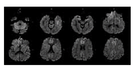 Diffusion weighted imaging
By measuring and imaging the difference in molecular diffusion in vivo, it is possible to diagnose an early brain stroke within 3 hours and to discern tissue abscess, tissue necrosis, and tumor.
-
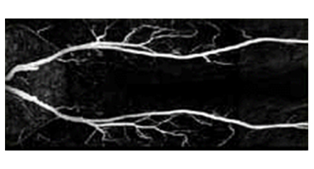
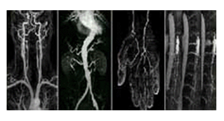 Magnetic resonance angiogram
Using a special imaging technique, even microscopic blood vessels in the human body can be presented in the form of an image.
-
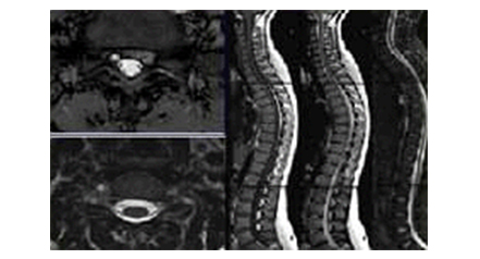 Spine image
By providing sharper images than the conventional MRI equipment, it provides excellent images for the diagnosis of disc herniation occurring in the spine.
-
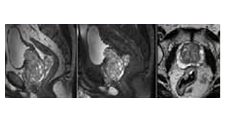 Prostate/Cervix Image
Using a special imaging technique, it can provide useful information in diagnosing prostate cancer or cervical cancer.
-
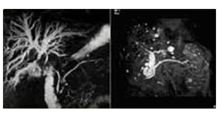
 Whole Body Angiogram
Using a special imaging technique, all the blood vessels in the human body can be presented in the form of an image.
-
 MRCP
MRCPThe biliary tract and pancreatic duct are scanned by optimizing the high signal strength of the pancreatic duct fluid or bile and the signal difference with surrounding tissues by using an imaging technique that emphasizes the T2 effect. It provides especially useful information for examining the upper biliary tract in the area where there is an obstruction.