Medical Equipment
OCT is a useful device for diagnosis and treatment of retinal diseases and glaucoma. Using a laser beam, it produces cross-sectional images of the retina and optic nerve that cannot be seen with the naked eye.
It enables diagnosis of retinal diseases that are difficult to identify with the naked eye or with conventional equipment by producing high-resolution images of the cross-sections of macula tissue, the most important part of the retina.
In addition, cross-sectional imaging of the micro-structures of the optic nerve enables an early detection of glaucoma and an examination of disease progression and treatment effects.
It is also possible to accurately measure the thickness of the retinal nerve fiber layer to determine the degree of nerve damage caused by glaucoma and the progression of the disease.
One of the benefits of using OCT is that patients experience no pain or discomfort at all.
1. cSLO Method
Sharper images are produced compared to the camera or SLO method
2. Auto Real Time (ART)
High-resolution images are produced by automatically averaging up to 100 frames within a short time.

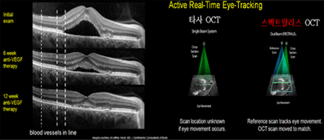
3. Eye Tracking
It is a dual beam system used to measure minute changes in lesions down to 1 μm.
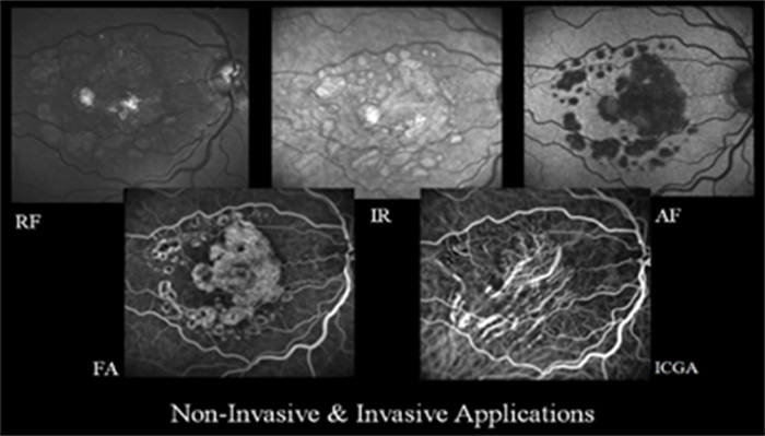
4. Various Shooting Modes
It shoots in various modes such as IR+OCT, FA+OCT, RF+OCT, ICGA+OCT, and AF+OCT simultaneously
5. High-SpeedScan Time
Spectralis HRA can capture up to 20 frames per second
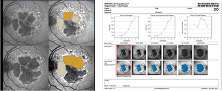
6. Regionfinder Software
It generates data on the changes in the atrophic area so that the changes can be checked over time
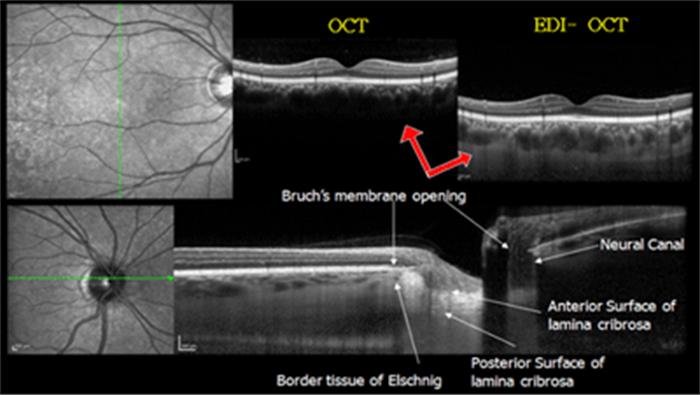
7. Enhanced Depth Imaging (EDI) Mode
Spectralis can be used to check for choroidis and examine new blood vessels forming in the choroid. When the optic nerve is photographed in EDI mode, the lamina cribrosa can be clearly observed, which can facilitate the diagnosis of glaucoma.
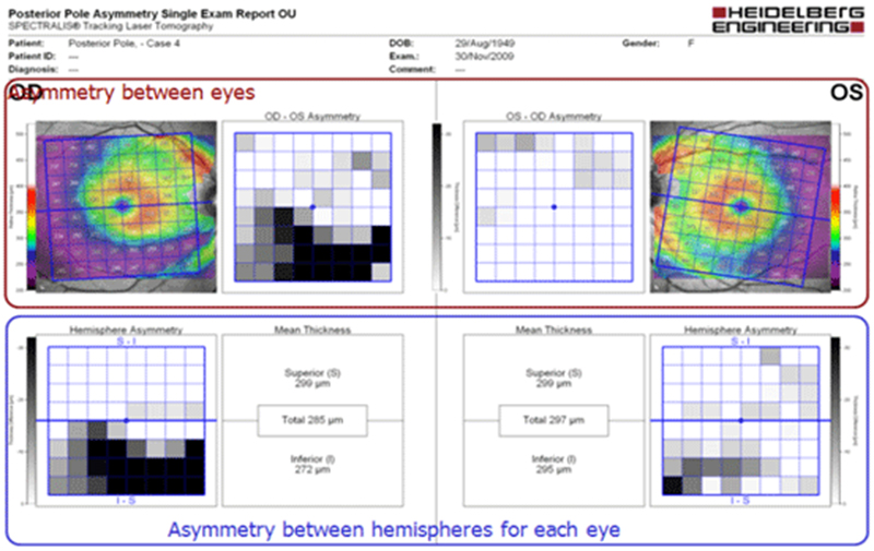
8. Posterior Pole Analysis
By measuring the thickness of the entire retina, the asymmetry between the eyes or monocular thickness of the ganglion cell layer can be determined for early detection of glaucoma
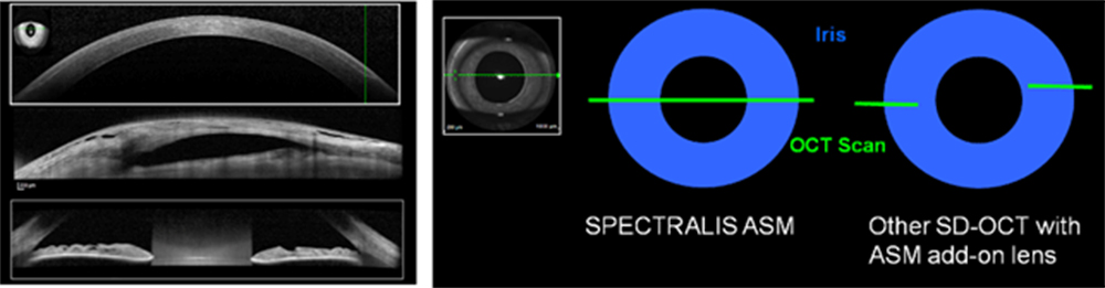
9. Anterior Segment Module
It can be used to acquire high-resolution 16mm images to diagnose LASIK flap as well as various anterior eye diseases